3D organoids in OMĒOS: from the freezer slot to the size table
Grow, image and measure organoids in one place: from the freezer slot and the passaging protocol to segmentation and size measurements.
What you can do in OMĒOS today
- Outline organoids in brightfield images with SAM 2, no training needed
- Measure organoid size image by image, and export the table
- Track organoid lines down to the freezer slot
- Watch time-lapses of organoids growing, in place
Coming soon: 3D cell labelling through light-sheet and confocal stacks.
See it in OMĒOS
- Segment organoids with SAM 2, an OMĒOS Short
- Measure what matters, an OMĒOS Short
- Track samples and reagents, an OMĒOS Short
Read more
- The logistical labyrinth: shipping live organ-on-a-chip systems and organoids
- Overcoming the challenges in portable cell culture incubation
Ready to try it? Download OMĒOS or watch all OMĒOS Shorts.
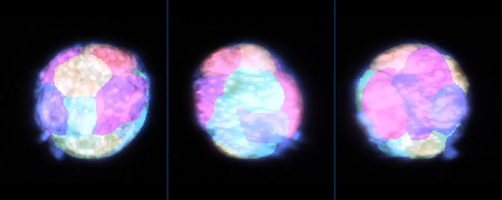
Mouse mammary organoid, dual oblique plane light-sheet; cell labels from the dataset’s authors. Smith MB, et al., Zenodo 2023, doi:10.5281/zenodo.7544194 (CC BY 4.0).
Tags