OMĒOS software

Your whole lab, on one screen.

Instruments, data, protocols and your team in one workspace, with AI that runs on your own machines.

  • Data scattered across instrument PCs and inboxesOne searchable tree
  • Analysis that lives on one person's laptopModels the whole team can run
  • Protocols lost in binders and old versionsLive, reviewed documents
The ON series

And everything ON it.

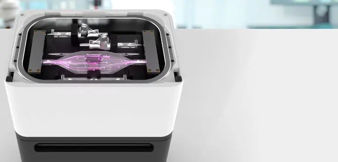
Hardware built to run with OMĒOS, from the bench to the cloud.

All products →
Coming soon

ON Edge

AI compute that stays in your lab, set up in minutes and run from OMĒOS.

SdeskMbenchXrack
Explore ON Edge →

30-day full refund1-week replacementsPrices on every page

Image and data credits

  • Organoid images: Hof L., Moreth T., Koch M., Pampaloni F., et al., BMC Biology 2021; data doi:10.5281/zenodo.4419985 (CC BY 4.0), cropped and adjusted.
  • HeLa time-lapse: R. Guiet, BioImaging and Optics Platform (BIOP), EPFL; doi:10.5281/zenodo.4700067 (CC BY 4.0), cropped and adjusted.
  • Organoid single-cell data: Moerkens et al., Cell Reports 2024 (doi:10.1016/j.celrep.2024.114247), via CZ CELLxGENE Discover (CC BY 4.0), subsampled.
  • H&E slide: OpenSlide test data (CMU-1), Carnegie Mellon University (CC0).
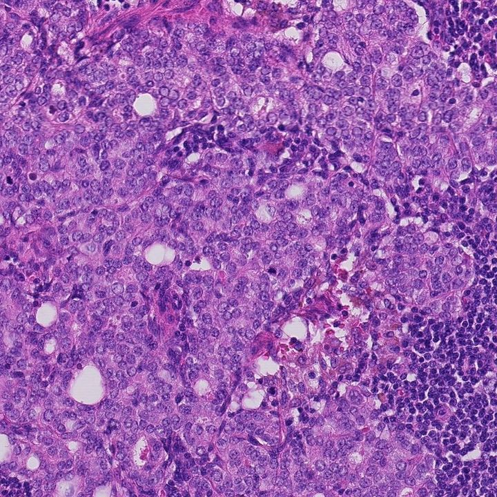
  • Lymph node H&E: CAMELYON16, Litjens G., et al., GigaScience 2018, via the Registry of Open Data on AWS (CC0).
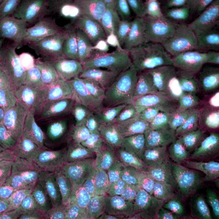
  • Cell Painting: JUMP cpg0016, Chandrasekaran et al. 2023; Cell Painting Gallery, Weisbart et al., Nature Methods 2024 (CC0).
  • Mammary organoid light-sheet: Smith M.B., et al., Zenodo 2023, doi:10.5281/zenodo.7544194 (CC BY 4.0).

Demo people and organizations in our videos are fictional. Scientific images are real open data, credited above.