Cellular microscopy in OMĒOS: segment and measure every cell
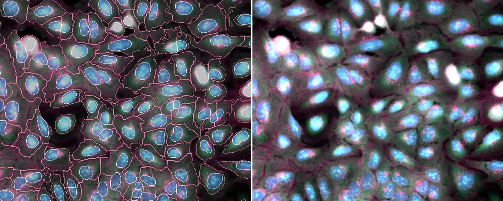
Bring multi-channel fluorescence into the same workspace as your protocols and data, segment cells and nuclei, and measure them image by image.
What you can do in OMĒOS today
- Open fluorescence images and time-lapse movies right where you work
- Segment cells and nuclei with models from the catalog, or your own
- Measure area, intensity and shape, and copy the table into a workbook
- Keep images, results and the protocol together in one project
Supported formats: CZI · ND2 · LIF · OME-TIFF.
See it in OMĒOS
- Measure what matters, an OMĒOS Short
- Find nuclei with StarDist, an OMĒOS Short
- Workbooks and databases, an OMĒOS Short
Read more
- Microscopy Resolution & Nyquist Calculator: optimal pixel size from NA & wavelength
- Open-source cellular image analysis in 2026: a field map of the tools that matter
Ready to try it? Download OMĒOS or watch all OMĒOS Shorts.
U2OS cells, Cell Painting (DNA, ER, actin/Golgi). JUMP cpg0016: Chandrasekaran et al. 2023; Cell Painting Gallery, Weisbart et al., Nature Methods 2024 (CC0). Outlines drawn by 37degrees.