Spatial biology in OMĒOS: keep every molecule with its cells
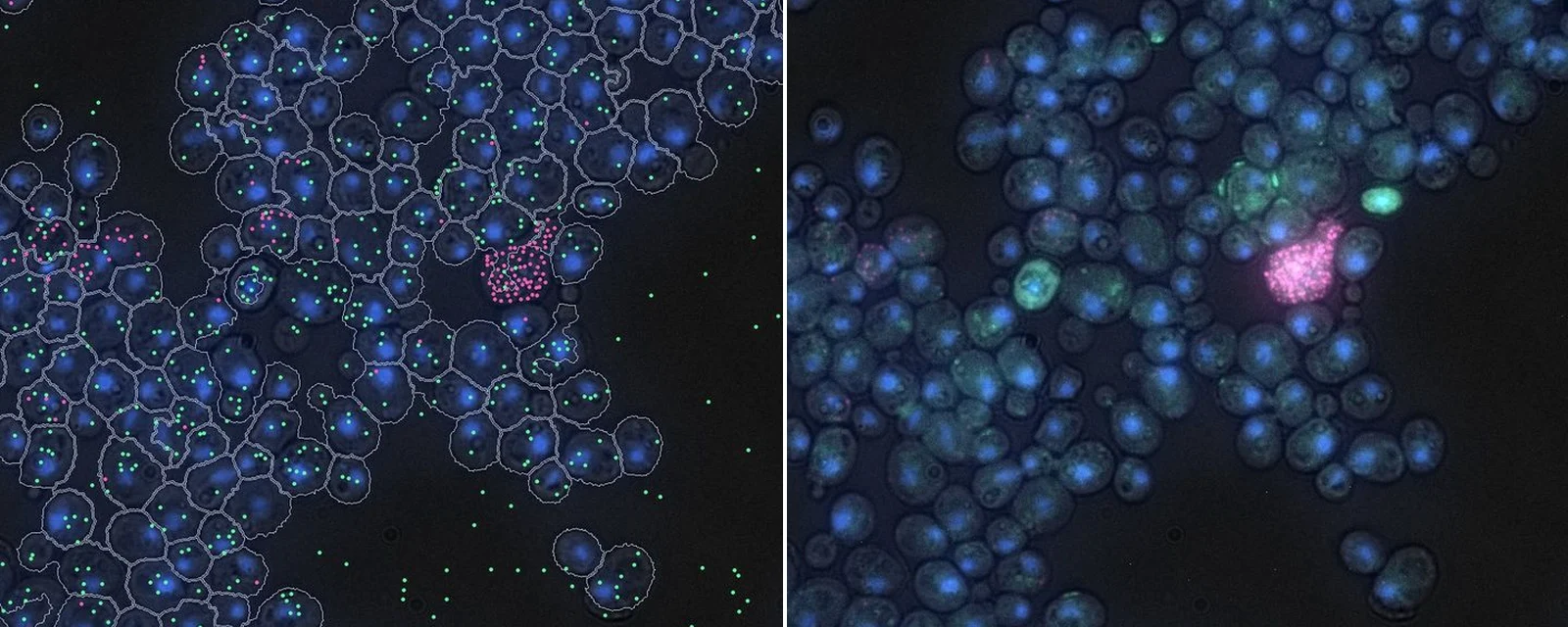
Keep single-molecule imaging next to the cells it came from, the sample record and the protocol, so every count can be traced back.
What you can do in OMĒOS today
- Open multi-channel images in place, channel by channel
- Segment the cells and measure each channel’s signal
- Link images to the sample in inventory and the protocol that made them
Coming soon: Automatic spot detection, and assigning each molecule to its cell.
See it in OMĒOS
- Open slides, movies and tables, an OMĒOS Short
- Track samples and reagents, an OMĒOS Short
- Measure what matters, an OMĒOS Short
Read more
Ready to try it? Download OMĒOS or watch all OMĒOS Shorts.
Yeast single-molecule RNA FISH, two transcripts. IDR idr0047 (Neuert lab); Image Data Resource, Williams E, et al., Nature Methods 14:775, 2017 (CC BY 4.0). Spots and outlines drawn by 37degrees.