NIH awards 37degrees, Inc. an STTR Phase I for PamON — longitudinal 3D imaging of tissue constructs — in partnership with the Pramanik Lab
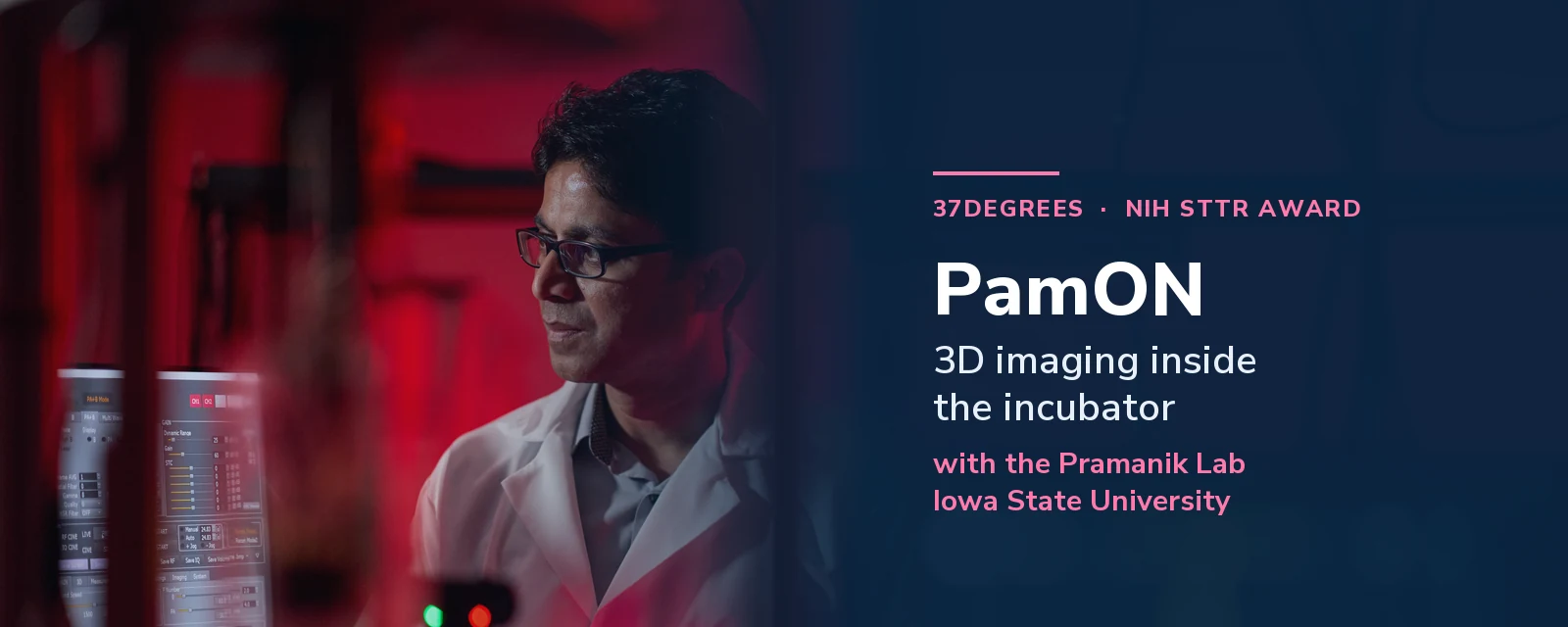
The National Institute of General Medical Sciences (NIGMS), part of the National Institutes of Health, has awarded 37degrees, Inc. a Small Business Technology Transfer (STTR) Phase I grant — $360,247 over twelve months, beginning 1 September 2026 — to develop PamON, a system for imaging living engineered tissue in three dimensions without taking it out of the incubator and without destroying it.
The optical engineering at the heart of the project is being carried out in collaboration with the Biomedical Imaging Laboratory of Dr. Manojit Pramanik, PhD at Iowa State University, who serves as research partner principal investigator. Dr. Tilak Jain, PhD, founder and CEO of 37degrees, is the principal investigator on the award.
The problem: you have to destroy the tissue to find out how it is doing
Engineered tissue — skin, cardiac and bone patches, organoids, spheroid co-cultures — is one of the more promising routes out of the organ shortage. Over 100,000 patients sit on the United States transplant waiting list; roughly 48,000 transplants were performed in 2024 [1, 2]. Tissue-engineered medical products won’t close that gap on their own, but they are already reshaping how repair, drug testing and disease modeling are done.
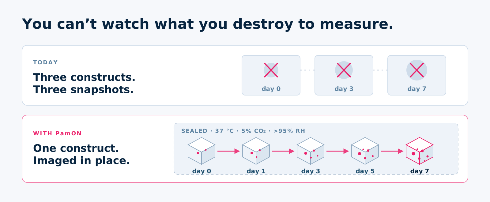
There is an awkward gap in the middle of that work. To find out whether a construct is maturing properly — whether cells are where they should be, whether it is vascularizing, whether the core is still alive — you generally have to fix it, section it, and stain it. The measurement consumes the sample [3, 4].
That has two consequences. You never see a trajectory, only a series of snapshots taken from different constructs. And a construct destined for a patient cannot be checked before it is released, because checking it is what destroys it.
This is not a niche complaint. A 2024 NIST workshop report (Special Publication 1500-23) gathered 180+ participants around exactly this question and came back with a unanimous call for non-destructive viability and functional characterization [5]. Independently, in an NSF I-Corps program, our team interviewed more than 150 scientists working with live tissue cultures — and the need to characterize tissue sections beyond what optical methods can reach came back as one of the top three findings.
Light goes in, sound comes out
The way around this is a technique called photoacoustic microscopy (PAM) [6, 7].
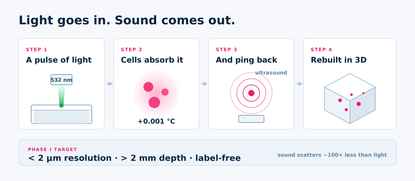
A very short pulse of laser light — a few billionths of a second — goes into the tissue. Whatever absorbs it (melanin, hemoglobin, nuclei, cytoskeleton) warms by a few thousandths of a degree, expands very slightly, and snaps back. That tiny expansion emits an ultrasound pulse. A fiber-optic sensor under the dish listens for it [13], and the arrival times are reconstructed into a 3D map of what absorbed the light and where [8].
The reason this reaches deeper than a confocal or multiphoton microscope is the return trip. Light scatters heavily on the way out of tissue; sound scatters roughly a hundred times less. So the signal that carries the depth information is the one that survives. And because the contrast comes from what the tissue already absorbs, no dye or fluorescent label is needed — which matters a great deal if the construct is eventually going into a patient. Label-free photoacoustic imaging has already been used to follow cells inside 3D porous scaffolds over weeks [9], and to track the same living tissue repeatedly over time [10].
What PamON actually is
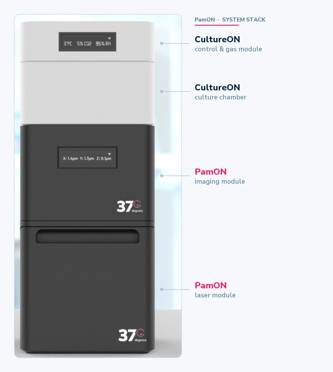
PamON is the photoacoustic module joined to the live-culture module — the two halves of the problem solved together.
The incubation half already exists. CultureON 100 is our portable CO₂ incubator: temperature, humidity and CO₂ control for long-term culture, running on pre-packaged CO₂ cartridges rather than a wall-mounted gas line [11]. For this project it carries a sealed optical port in the floor of the chamber — a window the imaging module can look through with the chamber staying closed, for minimum disturbance.
The imaging half is what this award funds. An optical-resolution photoacoustic microscope (OR-PAM) sits beneath that port in an inverted configuration, looking up through the window at the construct. In Phase I we are targeting a scan volume of roughly 1 cm² by 2 mm thick — enough for the research-scale tissue patches most groups work with.
Because the chamber stays closed and the construct is never touched, the same sample can be imaged again and again — the project targets seven days or more of repeat imaging on a single living construct, producing a genuine time-lapse of structural change rather than a set of unrelated snapshots.
Spotlight: the Biomedical Imaging Laboratory at Iowa State
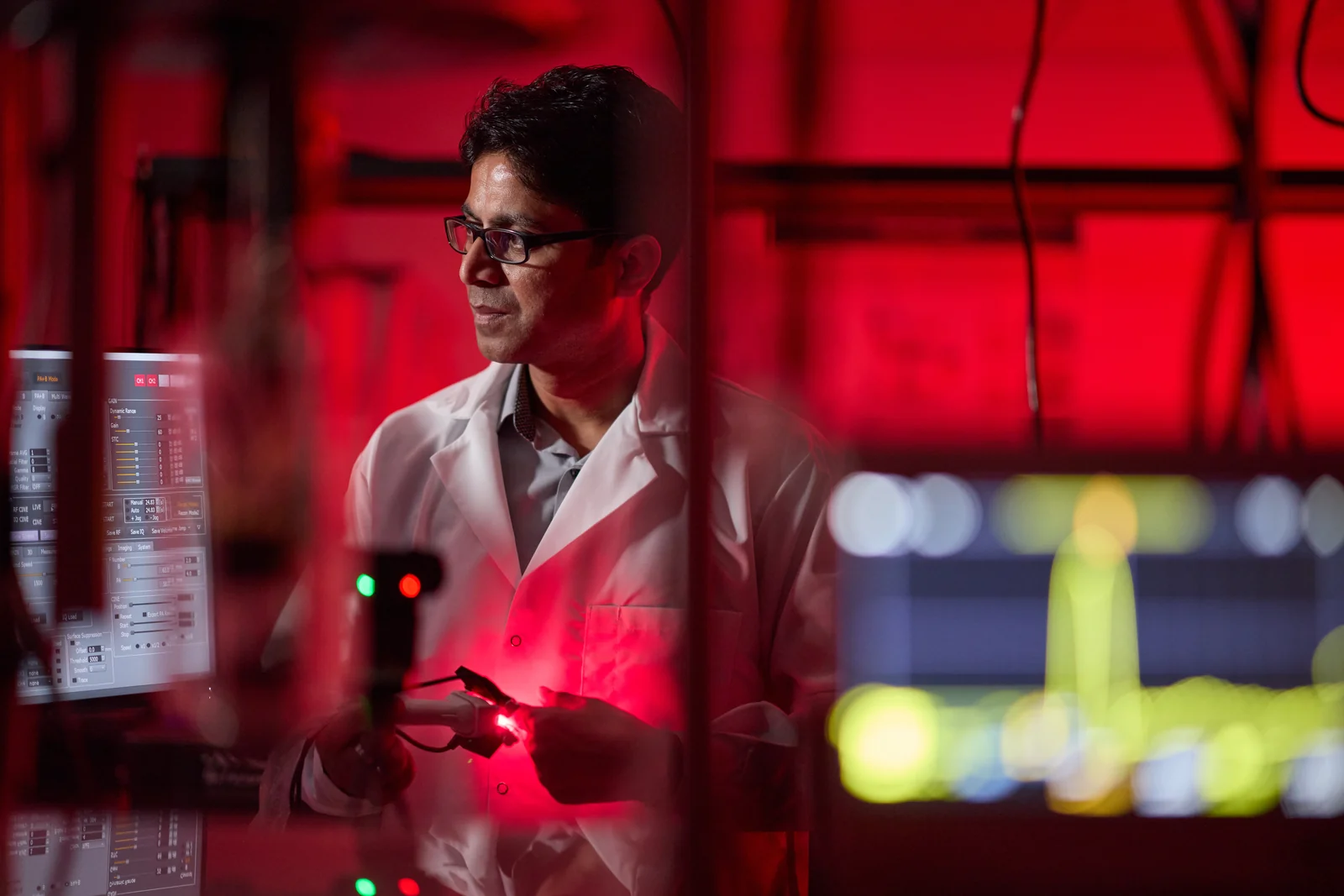
Dr. Manojit Pramanik, PhD is the Northrop Grumman Associate Professor in the Department of Electrical and Computer Engineering at Iowa State University, where he also holds an affiliated appointment in Biomedical Engineering. He runs the Biomedical Imaging Laboratory (BILab).
He came to photoacoustics early and has stayed with it for more than two decades. He earned his PhD in biomedical engineering at Washington University in St. Louis, in the environment where much of modern photoacoustic tomography was first built. Before academia he spent two years at GE Global Research and a year at Philips Medical Systems, both in Bangalore — an instrumentation background that shows in how his lab works. From 2014 to 2022 he was on the faculty at Nanyang Technological University in Singapore, joining Iowa State in 2023.
BILab’s work spans:
- Medical imaging systems — photoacoustic and thermoacoustic imaging, from benchtop microscopes to whole-organ tomography.
- Image reconstruction and processing, including machine-learning approaches to reconstruction and denoising.
- Contrast agents and molecular imaging.
- Biomedical device design — building the instruments, not only using them.
Clinically, the lab’s application areas run through breast cancer imaging, brain imaging, ovarian and pancreatic cancer, diabetes, and treatment monitoring.
Why this lab, for this problem
The specific prior result that makes PamON credible is a switchable acoustic-resolution / optical-resolution photoacoustic microscope the Pramanik lab built and published. In its optical-resolution mode it achieved 4.2 µm lateral resolution to a depth of 1.4 mm — fine enough to resolve single capillaries and cellular detail. Switched to acoustic-resolution mode, the same instrument reached 7.6 mm deep at 45 µm resolution [12].
That is the hard part of PamON already demonstrated, on the bench, by the people who will be building it. What is new here is putting an instrument of that class underneath a living culture that has to stay at 37 °C, 5% CO₂ and >95% humidity for a week or more — and getting it to hold micron-scale resolution while it does.
What comes next
Phase I runs to 31 August 2027. In parallel with the engineering, we are continuing customer discovery, early regulatory analysis for clinical applications, and beta-test planning with the tissue-engineering groups who have supported the project. Both teams intend to pursue an STTR Phase II application together.
If you work with thick 3D constructs, organoids, or tissue patches and the characterization step is costing you samples, we would like to hear how you work — that feedback is what shapes version 1.0.
- Contact 37degrees → — messages route directly to our team and are triaged within one business day.
- CultureON 100 → — the portable CO₂ incubator PamON is built around.
- OMĒOS → — the data platform where project datasets live.
References
- Organ Procurement and Transplantation Network (OPTN). Organ transplants exceeded 48,000 in 2024; a 3.3 percent increase from the transplants performed in 2023. U.S. Department of Health and Human Services, Health Resources and Services Administration, 2025.
- Lewis A, Koukoura A, Tsianos GI, et al. Organ donation in the US and Europe — the supply vs demand imbalance. Transplantation Reviews. 2021;35(2):100585.
- Zuncheddu D, Della Bella E, Schwab A, et al. Quality control methods in musculoskeletal tissue engineering — from imaging to biosensors. Bone Research. 2021;9(1):46. [PMC →]
- Appel AA, Anastasio MA, Larson JC, Brey EM. Imaging challenges in biomaterials and tissue engineering. Biomaterials. 2013;34(28):6615–6630. [PMC →]
- Babakhanova G, Simon CG Jr, Romantseva E. Measurement Needs for Biofabrication of Tissue Engineered Medical Products Workshop Report. NIST Special Publication 1500-23. U.S. Department of Commerce, National Institute of Standards and Technology, 2024. [NIST →]
- Cai X, Zhang YS, Xia Y, Wang LV. Photoacoustic microscopy in tissue engineering. Materials Today. 2013;16(3):67–77. [PMC →]
- Shrestha B, DeLuna F, Anastasio MA, Ye JY, Brey EM. Photoacoustic imaging in tissue engineering and regenerative medicine. Tissue Engineering Part B: Reviews. 2020;26(1):79–102. [PMC →]
- Wang LV, Yao J. A practical guide to photoacoustic tomography in the life sciences. Nature Methods. 2016;13(8):627–638. [PMC →]
- Zhang Y, Cai X, Choi SW, Kim C, Wang LV, Xia Y. Chronic label-free volumetric photoacoustic microscopy of melanoma cells in three-dimensional porous scaffolds. Biomaterials. 2010;31(33):8651–8658. [PMC →]
- Sciortino VM, Tran A, Sun N, et al. Longitudinal cortex-wide monitoring of cerebral hemodynamics and oxygen metabolism in awake mice using multi-parametric photoacoustic microscopy. Journal of Cerebral Blood Flow and Metabolism. 2021;41(12):3187–3199. [PMC →]
- Zhu AG, Reid E, Jain T, Mir A, Siddiqi U, Dunne O, Hibino N. Advancing tissue engineering through a portable perfusion and incubation system. Bioengineering. 2025;12(5):554. [PMC →] · [our summary →]
- Moothanchery M, Pramanik M. Performance characterization of a switchable acoustic and optical resolution photoacoustic microscopy system. Sensors. 2017;17(2):357. [PMC →]
- Guggenheim JA, Li J, Allen TJ, et al. Ultrasensitive plano-concave optical microresonators for ultrasound sensing. Nature Photonics. 2017;11(11):714.
Award information
PamON: 3D imaging of live and incubated engineered tissue with photoacoustic microscopy. NIH/NIGMS STTR Phase I, award number 1R41GM165131-01 (FAIN R41GM165131). Project period 1 September 2026 – 31 August 2027. Principal investigator: Tilak Jain, PhD, 37degrees, Inc. Research partner principal investigator: Manojit Pramanik, PhD, Iowa State University.
Research reported in this publication was supported by the National Institute of General Medical Sciences of the National Institutes of Health under Award Number R41GM165131. The content is solely the responsibility of the authors and does not necessarily represent the official views of the National Institutes of Health.
Learn more about the NIH SBIR/STTR programs →
About 37degrees
37degrees, Inc. designs portable, connected scientific instruments for life-science and biomedical research. The current instrument family addresses continuous live-cell culture and live-cell imaging, paired with the OMĒOS data platform; the broader product roadmap is an expanding instrument base spanning molecular and cellular research through translational and pre-clinical applications. The company is supported by the NSF SBIR program, the NVIDIA Inception Program, and AWS Activate.
Frequently asked questions
- What is PamON?
- PamON is a photoacoustic microscope being built to attach to a live-culture incubation module, so a 3D tissue construct can be imaged in three dimensions while it stays alive at 37 °C, 5% CO₂ and >95% humidity. It is designed to replace the destructive fix-section-stain step that engineered tissue characterization depends on today.
- How is photoacoustic microscopy different from confocal or multiphoton microscopy?
- Fluorescence methods send light in and read light back out, and light scatters heavily in tissue — which is why they typically run out of usable signal below about 1 mm and usually need a dye or a fluorescent label. Photoacoustic microscopy sends light in but reads ultrasound back out. Sound scatters roughly a hundred times less than light in tissue, so the return signal survives from deeper in the sample, and it works on what the tissue already absorbs — no label required.
- Who is funding this work?
- The National Institute of General Medical Sciences, part of the National Institutes of Health, under STTR Phase I award 1R41GM165131-01. The project period runs 1 September 2026 to 31 August 2027.
- What is an STTR award, and how does it differ from SBIR?
- Both are federal small-business research programs. The difference is that STTR requires the small business to formally partner with a non-profit research institution and to subaward a substantial share of the work to it. For PamON, that research partner is Dr. Manojit Pramanik's laboratory at Iowa State University.
- Can I buy PamON?
- Not yet. Phase I is a twelve-month feasibility project to build and characterize a first prototype. Research groups interested in beta testing a later version, or in the CultureON 100 incubator PamON is built around, are welcome to get in touch.