NIH awards 37degrees, Inc. an SBIR Phase I for InVitreON — long-term recording and stimulation of living neurons — in collaboration with Salk Institute scientist Rusty Gage†
The National Institute of Mental Health (NIMH), part of the National Institutes of Health, has awarded 37degrees, Inc. a Small Business Innovation Research (SBIR) Phase I grant over two years, beginning 1 August 2026, to develop InVitreON. InVitreON is a compact, integrated platform for growing living neurons, recording their electrical activity and stimulating them in closed loop for weeks at a time, without ever opening the chamber.
The neurobiology for the project is being carried out in collaboration with the Laboratory of Genetics of Dr. Rusty Gage at the Salk Institute for Biological Studies†. Dr. Melissa Hernandez, Senior Research Associate in Gage’s lab, leads the Salk side of the work. Dr. Tilak Jain, PhD, founder and CEO of 37degrees, is the principal investigator on the award.
† The Salk Institute for Biological Studies is participating in this project solely as a research collaborator, under a subaward from 37degrees, Inc. The Salk Institute, the Gage Laboratory and their investigators do not endorse, recommend or promote InVitreON, CultureON 100, OMĒOS or any other product or service of 37degrees, Inc. References to the Salk Institute and its scientists in this article describe their role in the research only and should not be read as an endorsement.
The problem: a powerful tool that most labs can’t easily use
Nervous system disorders are now the leading cause of illness worldwide, affecting an estimated 3.4 billion people, or 43% of the world’s population [1]. Many of the best tools for understanding them, and for finding drugs that work, are living neurons studied outside the body: primary rodent neurons, human iPSC-derived neurons, 3D brain organoids and organotypic brain slices.
For almost fifty years the standard way to listen to these ex-vivo neurons has been the microelectrode array (MEA): a glass dish with a grid of electrodes in its floor [2–4]. Neurons grown on it “wire” themselves into a network, and every electrode hears the spikes of the cells above it. MEAs are used in basic neurophysiology, disease modeling, neurotoxicity screening and drug development [5, 6]. Yet they are nowhere near as common in neuroscience labs as the microscope. There are five reasons:
- The rig is a collection of separate instruments. A typical long-term MEA experiment pieces together a culture incubator on mains power and a gas tank, pre-amplifiers, a data-acquisition computer, a perfusion pump, and light and electrical stimulators. All of it is joined by cables and tubing across a bench, and the long cables pick up electrical noise [7].
- The culture conditions aren’t physioxic. Neurons in the body live at far lower oxygen than room air’s 21%, and the damage atmospheric oxygen does to cultured neurons has been documented for decades [8]. Almost every incubator controls CO₂ only.
- Every media change is a risk. Cultures that need to run for weeks or months must be carried to a hood for each media change. Every trip invites contamination and disturbs the network. Sealed, breathable culture lids help a great deal [9], but they have never been built into an MEA system at scale.
- Stimulation comes one mode at a time. Driving a network with light, current or drugs, especially in closed loop in response to what it is doing, is a well-established, powerful technique [10–12]. But no single system brings chemical, optical and electrical stimulation together with the culture environment. Combining them takes a lab with deep engineering support [7].
- You can’t watch the same cells over time. Imaging a culture usually means taking it out of the incubator and off the rig. Pairing a cell’s electrical activity with what it looks like, week after week, is rarely possible.
What InVitreON is
InVitreON grows out of CultureON 100, our portable CO₂ incubator. CultureON 100 already provides temperature, humidity and CO₂ control on pre-packaged gas cartridges and battery power, with no gas line and no wall socket required [13]. InVitreON builds on that base and adds everything a long-term neuronal experiment needs:
- Physioxic oxygen levels. A second, nitrogen cartridge pushes oxygen in the chamber down to physiological levels. The chamber settles at 37 °C, 5% CO₂ and low O₂.
- Recording built in. The culture dish sits on the electrode array, and built-in pre-amplifiers connect through spring contacts the moment the lid closes, keeping the signal path short and quiet.
- A sealed, breathable cap. The dish is closed with a gas-permeable membrane, so the network stays sterile and its media doesn’t concentrate as it evaporates [9].
- Fluidics while recording. A cooled drawer holds fresh media and test compounds. A gentle, low-flow pump exchanges them through ports in the cap without interrupting the recording.
- Three ways to stimulate. Light, to excite or inhibit optogenetically modified neurons; current pulses through the electrodes; and compounds delivered through the media line.
- Compute on board. An embedded computer records the electrodes, sorts the spikes and runs closed-loop rules in real time. Data streams to OMĒOS, our cloud software, for remote monitoring and control of the experiment.
- A window underneath. The glass array is transparent, so the same neurons can be imaged while they are being recorded, for example with a calcium indicator [14].
Using it is meant to be simple. You load the gas, media and reagent cartridges, put in a culture dish with its sealed cap, and close the lid. Closing it makes the electrical and fluidic connections. You then set the environment, recording and stimulation in software and leave the network to grow. Because the unit runs on a battery, it can be moved without stopping a recording.
Listening, and talking back
An MEA tells you what a network is doing. The more interesting experiments start when you can answer it. Feeding a network stimuli that depend on its own activity has been used to control bursting in cortical cultures [10], to steer networks with light [11], and to study learning in living circuits [12].
In InVitreON all three kinds of stimulus are in the box and run by the same computer that is reading the electrodes. A rule like “when this region bursts, inhibit it with light” or “pulse here whenever that electrode fires” is a software setting, not a week of wiring.
What Phase I will test
The first experiments are deliberately standard, chosen to prove the platform against well-known results:
- Environment. Rodent cortical and hippocampal neurons, plus human iPSC-derived glutamatergic and dopaminergic neurons, are grown at a range of oxygen levels and compared with cultures in a conventional incubator.
- Recording. Networks are recorded for one to two months, with media exchanged automatically, and compared with the firing and bursting patterns reported in the literature [15].
- Stimulation and imaging. Optogenetic excitation and silencing, electrical stimulation and drug application are used on the same cultures, with calcium imaging alongside the electrode recordings.
These are first tests, not the limit of the platform. InVitreON is designed as a general tool for neuroscience, including:
- Disease modeling. Neurodevelopmental, neurodegenerative and psychiatric conditions, in patient-derived human iPSC neurons.
- Neurotoxicity and drug screening. Where network activity is a sensitive, functional readout [16]. Human-cell assays like these are part of the new approach methodologies (NAMs) that regulators are encouraging as alternatives to animal testing.
- 3D brain organoids, neural spheroids and organotypic brain slices. These are complex models that need to be kept healthy and followed for weeks rather than hours.
- Closed-loop and learning studies of how living networks adapt.
- Neural interface research. Working out how to read from and write to living neurons, groundwork for brain–machine interfaces.
Spotlight: Dr. Rusty Gage’s Laboratory at the Salk Institute

Dr. Rusty Gage at the Salk Institute for Biological Studies. Photo: Janet Bark / Salk Institute.
Dr. Rusty Gage is a Professor at the Salk Institute, where he holds the Vi and John Adler Chair for Research on Age-Related Neurodegenerative Disease. He served as President of the Salk Institute from 2018 to 2023. He is a member of the National Academy of Sciences, the National Academy of Medicine, the American Academy of Arts and Sciences and the American Philosophical Society. He has served as president of both the Society for Neuroscience and the International Society for Stem Cell Research. His recent honors include the 2024 Ogawa-Yamanaka Stem Cell Prize and the 2024 J. Allyn Taylor International Prize in Medicine.
His laboratory has changed how we think about the adult brain more than once:
- The adult human brain makes new neurons. In 1998, with colleagues in Sweden, the lab showed that neurogenesis continues in the adult human hippocampus. That overturned the long-held belief that we are born with all the neurons we will ever have [17].
- Experience shapes that growth. The lab showed that an enriched environment and physical exercise increase the birth of new neurons in the adult brain [18, 19].
- Neurons in the same brain can differ genetically. The lab found that “jumping genes” (LINE-1 retrotransposons) are active in developing neurons, creating genetic diversity from cell to cell within a single brain [20].
- Patients’ own neurons, in a dish. The Gage lab pioneered the use of iPSC-derived neurons, made from patients’ own cells, to study psychiatric illness. This includes a landmark early model of schizophrenia [21], and the discovery that neurons from people with bipolar disorder are hyperexcitable, with that hyperexcitability calmed by lithium only in neurons from patients who respond to the drug [22].
- Neurons that remember their age. The lab showed that neurons converted directly from skin cells keep the molecular signatures of the donor’s age, which opens the way to studying the aging brain in a dish [23].
- Human organoids, wired in. The lab grafted human 3D brain organoids into the mouse brain, where they developed blood vessels and functional connections with the host [24].
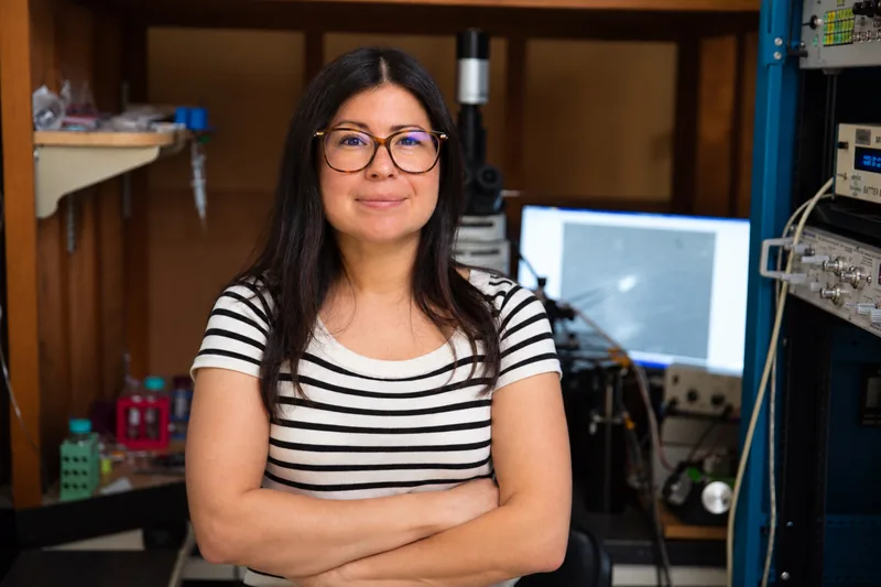
The Salk side of the InVitreON project is led by Dr. Melissa Hernandez, Senior Research Associate in Dr. Gage’s lab, who brings expertise in neuronal electrophysiology and human neuron models. Photo: Salk Institute.
Salk’s functional work on neurons
Much of the Gage lab’s most important work is functional: what patient-derived neurons do, measured electrically, over time. 37degrees will be working with Salk to validate InVitreON, harnessing the Gage lab’s functional expertise with neurons. The lab also brings well-established methods that the Phase I tests rely on directly, including lentiviral delivery of the optogenetic tools used to switch neurons on and off with light.
What comes next
Phase I runs to 31 July 2028. Engineering takes place at our Chicago facility and biological testing at Salk, with the two teams working back and forth between Chicago and La Jolla. In parallel we are continuing customer discovery with neuroscience groups and laying the groundwork for Phase II.
Photo credits
- The Salk courtyard, with Jonas Salk's words carved in the stone. : Chris Keeney / Salk Institute , © Salk Institute, used with permission
- Dr. Rusty Gage. : Salk Institute , © Salk Institute, used with permission
- Sunset over the Pacific from the Salk courtyard in La Jolla. : Chris Keeney / Salk Institute , © Salk Institute, used with permission
If you record from neurons on MEAs, or would like to but have been put off by the setup, we would like to hear how you work. That feedback is what shapes version 1.0.
- Contact 37degrees →: messages route directly to our team and are triaged within one business day.
- CultureON 100 →: the portable CO₂ incubator InVitreON builds on, available today.
- OMĒOS →: our cloud software for monitoring and controlling experiments remotely.
References
- GBD 2021 Nervous System Disorders Collaborators. Global, regional, and national burden of disorders affecting the nervous system, 1990–2021: a systematic analysis for the Global Burden of Disease Study 2021. Lancet Neurology. 2024;23(4):344–381. [PMC →]
- Thomas CA Jr, Springer PA, Loeb GE, Berwald-Netter Y, Okun LM. A miniature microelectrode array to monitor the bioelectric activity of cultured cells. Experimental Cell Research. 1972;74(1):61–66.
- Gross GW, Rieske E, Kreutzberg GW, Meyer A. A new fixed-array multi-microelectrode system designed for long-term monitoring of extracellular single unit neuronal activity in vitro. Neuroscience Letters. 1977;6(2–3):101–105.
- Pine J. Recording action potentials from cultured neurons with extracellular microcircuit electrodes. Journal of Neuroscience Methods. 1980;2(1):19–31.
- Cerina M, Piastra MC, Frega M. The potential of in vitro neuronal networks cultured on micro electrode arrays for biomedical research. Progress in Biomedical Engineering. 2023;5:032002. [DOI →]
- Chiappalone M, Pasquale V, Frega M (eds). In Vitro Neuronal Networks: From Culturing Methods to Neuro-Technological Applications. Springer; 2019.
- Zhang X, Dou Z, Kim SH, et al. Mind In Vitro platforms: versatile, scalable, robust, and open solutions to interfacing with living neurons. Advanced Science. 2024;11:e2306826. [PMC →]
- Brewer GJ, Cotman CW. Survival and growth of hippocampal neurons in defined medium at low density: advantages of a sandwich culture technique or low oxygen. Brain Research. 1989;494(1):65–74.
- Potter SM, DeMarse TB. A new approach to neural cell culture for long-term studies. Journal of Neuroscience Methods. 2001;110(1–2):17–24.
- Wagenaar DA, Madhavan R, Pine J, Potter SM. Controlling bursting in cortical cultures with closed-loop multi-electrode stimulation. Journal of Neuroscience. 2005;25(3):680–688. [PMC →]
- Newman JP, Fong M-F, Millard DC, et al. Optogenetic feedback control of neural activity. eLife. 2015;4:e07192. [PMC →]
- Bakkum DJ, Chao ZC, Potter SM. Spatio-temporal electrical stimuli shape behavior of an embodied cortical network in a goal-directed learning task. Journal of Neural Engineering. 2008;5(3):310–323. [PMC →]
- Zhu AG, Reid E, Jain T, Mir A, Siddiqi U, Dunne O, Hibino N. Advancing tissue engineering through a portable perfusion and incubation system. Bioengineering. 2025;12(5):554. [PMC →] · [our summary →]
- Shew WL, Bellay T, Plenz D. Simultaneous multi-electrode array recording and two-photon calcium imaging of neural activity. Journal of Neuroscience Methods. 2010;192(1):75–82. [PMC →]
- Wagenaar DA, Pine J, Potter SM. An extremely rich repertoire of bursting patterns during the development of cortical cultures. BMC Neuroscience. 2006;7:11. [PMC →]
- Johnstone AFM, Gross GW, Weiss DG, Schroeder OH-U, Gramowski A, Shafer TJ. Microelectrode arrays: a physiologically based neurotoxicity testing platform for the 21st century. NeuroToxicology. 2010;31(4):331–350.
- Eriksson PS, Perfilieva E, Björk-Eriksson T, Alborn AM, Nordborg C, Peterson DA, Gage FH. Neurogenesis in the adult human hippocampus. Nature Medicine. 1998;4(11):1313–1317. [DOI →]
- Kempermann G, Kuhn HG, Gage FH. More hippocampal neurons in adult mice living in an enriched environment. Nature. 1997;386(6624):493–495. [DOI →]
- van Praag H, Kempermann G, Gage FH. Running increases cell proliferation and neurogenesis in the adult mouse dentate gyrus. Nature Neuroscience. 1999;2(3):266–270. [DOI →]
- Muotri AR, Chu VT, Marchetto MC, Deng W, Moran JV, Gage FH. Somatic mosaicism in neuronal precursor cells mediated by L1 retrotransposition. Nature. 2005;435(7044):903–910. [DOI →]
- Brennand KJ, Simone A, Jou J, et al., Gage FH. Modelling schizophrenia using human induced pluripotent stem cells. Nature. 2011;473(7346):221–225. [PMC →]
- Mertens J, Wang QW, Kim Y, et al., Gage FH, Yao J. Differential responses to lithium in hyperexcitable neurons from patients with bipolar disorder. Nature. 2015;527(7576):95–99. [PMC →]
- Mertens J, Paquola ACM, Ku M, et al., Gage FH. Directly reprogrammed human neurons retain aging-associated transcriptomic signatures and reveal age-related nucleocytoplasmic defects. Cell Stem Cell. 2015;17(6):705–718. [PMC →]
- Mansour AA, Gonçalves JT, Bloyd CW, et al., Gage FH. An in vivo model of functional and vascularized human brain organoids. Nature Biotechnology. 2018;36(5):432–441. [PMC →]
Award information
InVitreON: A compact and integrated platform for long-term neuronal culture incubation, recording and closed-loop stimulation. NIH/NIMH SBIR Phase I, award number 1R43MH139167-01A1 (FAIN R43MH139167). Project period 1 August 2026 – 31 July 2028. Principal investigator: Tilak Jain, PhD, 37degrees, Inc. Subaward: Salk Institute for Biological Studies, led by Melissa Hernandez, PhD.
Funding: $456,902 awarded for year one, with $439,816 recommended for year two subject to the availability of funds and satisfactory progress, for a total of up to $896,718. The project is 100% federally funded ($896,718); 0% ($0) of total project costs is financed by non-governmental sources.
Research reported in this publication was supported by the National Institute of Mental Health of the National Institutes of Health under Award Number R43MH139167. The content is solely the responsibility of the authors and does not necessarily represent the official views of the National Institutes of Health.
Learn more about the NIH SBIR/STTR programs →
Photographs of the Salk Institute, Dr. Gage and Dr. Hernandez courtesy of the Salk Institute for Biological Studies. Banner photograph: Chris Keeney / Salk Institute.
About 37degrees
37degrees, Inc. designs portable, connected scientific instruments for life-science and biomedical research. The current instrument family addresses continuous live-cell culture and live-cell imaging, paired with the OMĒOS data platform; the broader product roadmap is an expanding instrument base spanning molecular and cellular research through translational and pre-clinical applications. The company is supported by the National Institutes of Health, the NSF SBIR program, the NVIDIA Inception Program, and AWS Activate.
Frequently asked questions
- What is InVitreON?
- InVitreON is a compact, battery-powered platform for long-term work with living neurons. It holds the culture at 37 °C with controlled CO₂ and oxygen, records the network's electrical activity on a microelectrode array, stimulates it with light, current or compounds, and exchanges media automatically — all with the chamber closed, for weeks at a time.
- What is a microelectrode array (MEA)?
- A glass dish with a grid of tiny electrodes built into its floor. Neurons grown on it form a living network, and each electrode picks up the spikes of the cells above it, so the activity of the whole network can be followed without touching a single cell. MEAs have been used since the 1970s in basic neurophysiology, disease modeling, neurotoxicity testing and drug development.
- Why control oxygen as well as CO₂?
- Neurons in the brain live at far lower oxygen than room air's 21%, and the toxic effect of atmospheric oxygen on cultured neurons has been documented for decades. Most incubators only control CO₂. InVitreON uses small nitrogen cartridges to bring oxygen down to physioxic levels.
- Who is funding this work?
- The National Institute of Mental Health, part of the National Institutes of Health, under SBIR Phase I award 1R43MH139167-01A1. The project period runs 1 August 2026 to 31 July 2028.
- Can InVitreON be used with brain organoids or organotypic brain slices?
- That is where we are headed. Phase I validates the platform on primary rodent neurons and human iPSC-derived neurons. 3D brain organoids, neural spheroids and organotypic brain slices are natural next models, because they need long, undisturbed culture at physioxic oxygen, which is exactly what InVitreON is designed to provide.
- Can I buy InVitreON?
- Not yet. Phase I is a feasibility project to build and test the first integrated system. Neuroscience groups interested in early access, or in the CultureON 100 incubator that InVitreON builds on, are welcome to get in touch. The Salk Institute is a research collaborator on the project only and does not sell, distribute or endorse 37degrees products.