How labs use OMĒOS, area by area.
November 17, 2026
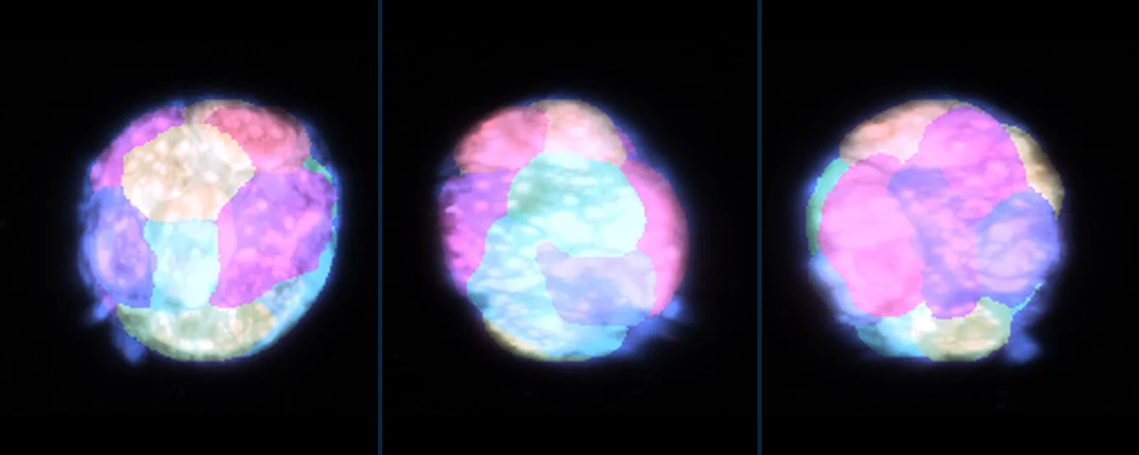
Grow, image and measure organoids in one place: from the freezer slot and the passaging protocol to segmentation and size measurements.
Read post →
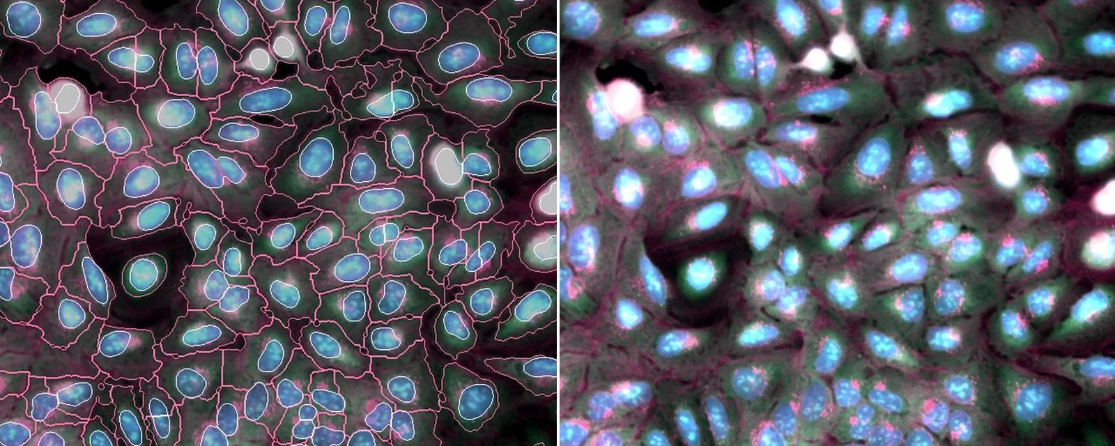
Bring multi-channel fluorescence into the same workspace as your protocols and data, segment cells and nuclei, and measure them image by image.
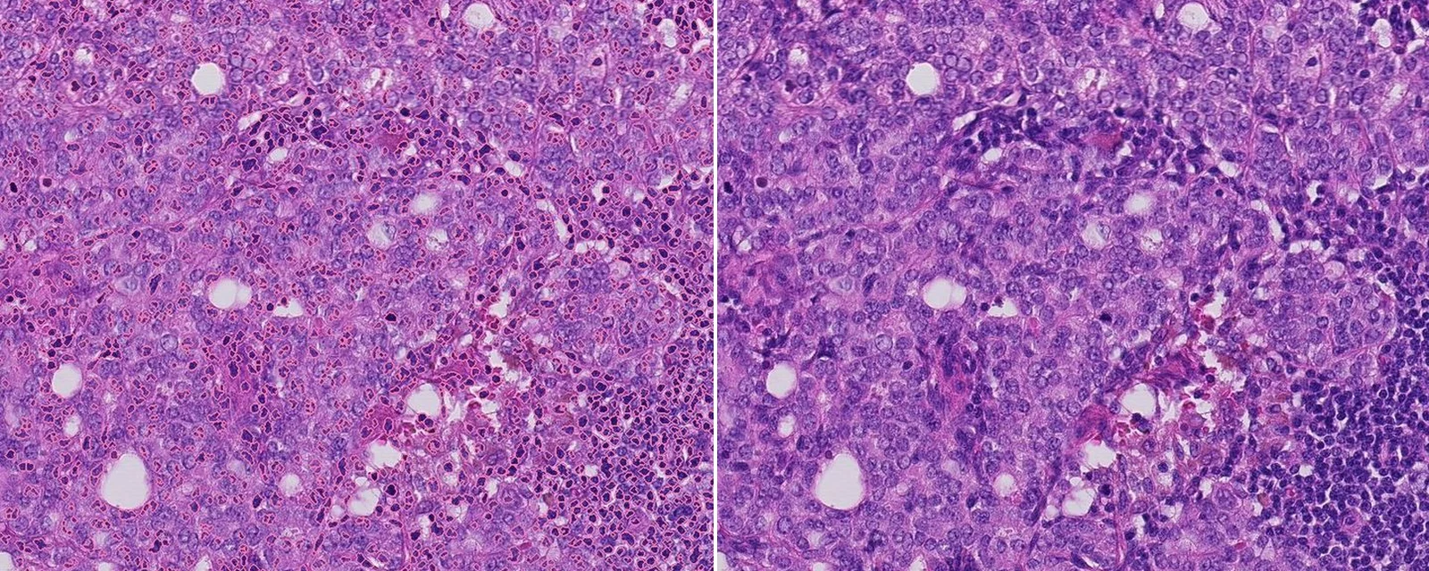
Open gigapixel slides on the desktop or in the browser, find every nucleus with a segmentation model running on your own machine, and share the result with your team.
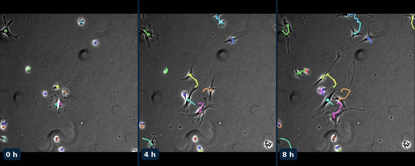
Keep brightfield and phase-contrast time-lapses with the experiment they belong to, play them in place, and analyse them frame by frame.
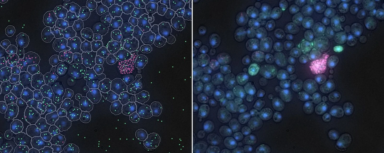
Keep single-molecule imaging next to the cells it came from, the sample record and the protocol, so every count can be traced back.
Subscribe
News, technical articles, research publications, and interactive tools — from 37degrees.
Unsubscribe anytime.
We use essential cookies to run the site. With your permission, we'd also like to use Google Analytics to understand how the site is used. See our cookie policy .